saxon Animals Biological Microscope Prepared Slides -50pcs
saxon Animal Biological Microscope Prepared Slides – 50 Piece Educational Set
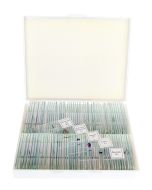
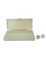
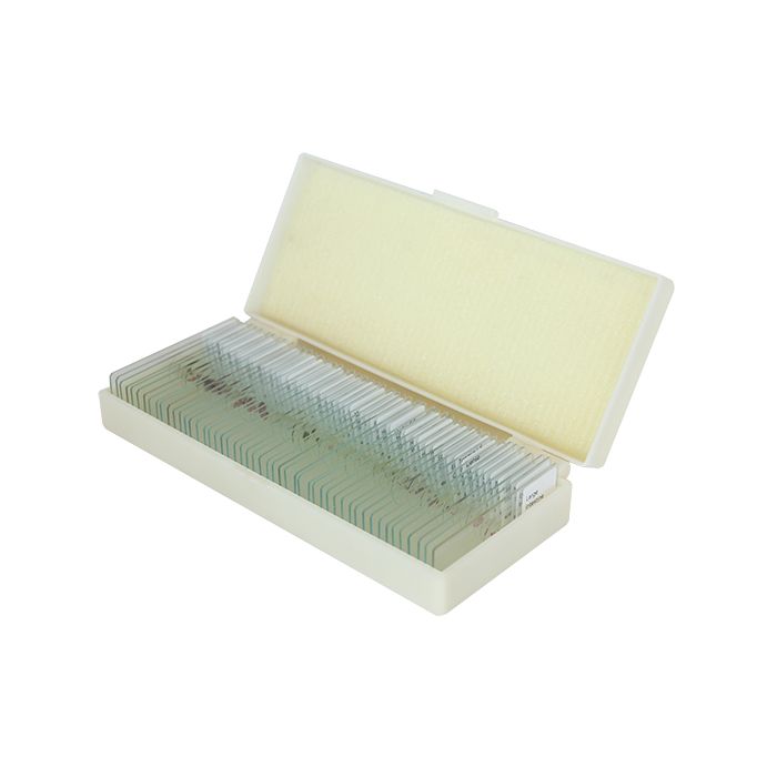
Explore the wonders of animal biology with the saxon 50 Piece Prepared Microscope Slide Set—a comprehensive educational resource designed for students, educators, and curious minds of all ages. This professionally curated collection features 50 labelled slides showcasing a wide variety of animal tissues, cells, and microscopic organisms.
Perfect for school labs, homeschooling, or personal exploration, this slide set offers a hands-on look into microscopic anatomy, organ systems, cell structures, and developmental stages. Each slide is safely housed in a protective hard carry case for secure storage and easy organization.
Slide Index
- Mouth Cell – Human cheek cell structure
- Single Ply Squamous Epithelium W.M – Thin, flat cells lining surfaces
- Stratified Squamous Epithelium Sec – Multi-layered protective tissue
- Skeletal Muscle L.S & C.S – Muscle fibers in longitudinal and cross-section
- Smooth Muscle C.S – Involuntary muscle tissue found in organs
- Cardiac Muscle L.S – Heart muscle with striations and intercalated discs
- Lung Vascular Injection Sec – Blood vessels in lung tissue
- Lymph Node Sec – Immune tissue structure
- Frog Blood Smear – Nucleated red blood cells
- Mammal Blood Smear – Red and white blood cells
- Stomach Paries Sec – Layers of stomach wall
- Frog Blastula Sag Sec – Early frog embryo development
- Human Hair Follicle Sec – Root structure of hair
- Artery and Vein Vascular C.S – Comparison of blood vessels
- Kidney L.S (Reins) – Kidney tubule structure
- Ovary Sec – Female reproductive organ
- Bladder Sec – Transitional epithelium in bladder wall
- Sperm Smear – Male reproductive cells
- Motor Nerve Cell W.M – Nerve cell with visible processes
- Tapeworm Mature Proglottid W.M – Reproductive section of tapeworm
- Frog Cleavage Sag Sec – Cell division in early embryo
- Honey Bee Mouth Parts W.M – Feeding structures of bee
- Butterfly Mouth Parts W.M – Coiled proboscis structure
- Housefly Mouth Parts W.M – Sponging mouth anatomy
- Female Mosquito Mouth Parts W.M – Piercing and feeding structures
- Male & Female Ascarid C.S – Parasitic worm anatomy
- Daphnia W.M – Tiny freshwater crustacean
- Hydra L.S – Longitudinal view of freshwater polyp
- Planaria W.M (injected digestive system) – Flatworm showing digestive tract
- Hydra Budding Whole W.M – Asexual reproduction in hydra
- Paramecium W.M – Ciliated protozoan organism
- Hydra C.S Through Spermary – Male reproductive tissue
- Hydra C.S Through Ovary – Female reproductive tissue
- Housefly Wing W.M – Transparent wing venation
- Butterfly Scale W.M (Squama) – Pigmented wing scale
- Housefly Foot W.M – Claw and pad structure
- Honey Bee Third Pair of Legs W.M – Pollen-carrying limb
- Spinal Cord C.S – Central nervous system anatomy
- Small Intestine Sec – Villi and epithelial lining
- Earthworm C.S – Internal body segmentation
- Tongue L.S (Cat or Dog) – Filiform papilla structure
- Dense Connective Tissue (Tendon L.S) – Parallel collagen fibers
- Loose Connective Tissue – Areolar tissue with fibers and cells
- Ciliated Epithelium Sec – Cells with hair-like cilia
- Frog Egg 2-Cell Sec – Embryo at 2-cell division stage
- Insect Compound Eye W.M – Ommatidia structure
- Taste Bud Sec (Rabbit) – Sensory cells for taste
- Nervous Tissue Sec – Neurons and support cells
- Eye Worm Tissue – Parasitic worm section
- Large Intestine Sec – Lumen and goblet cells
Educational and Engaging
Whether used in a classroom, laboratory, homeschool environment, or as a personal exploration tool, the saxon Animal Biological Prepared Slide Set offers an interactive and immersive way to learn biology through direct observation. This set brings science to life—slide by slide.
| Manufacturer | saxon |
|---|